Shoulder Muscles Diagram Posterior : Crossfit Shoulder Muscles Part 2 Posterior Musculature / Posterior part of the deltoid:. When a bilateral posterior dislocation is present, it is almost always secondary to seizure activity. Extends and laterally rotates the arm. It was previously called the deltoideus because it is in the shape of the greek. Infraspinatus and teres minor tendon. Medical illustration of the shoulder's muscles :
Learn vocabulary, terms and more with flashcards, games and other study tools. Start studying posterior shoulder muscles. The shoulder muscles are associated with movements of the upper limb. The shoulder muscles include skeletal muscles that are attached to the head of the humerus which performs various direct and indirect functions of the shoulder joints. With seizure activity, the internal rotator muscles (teres major.
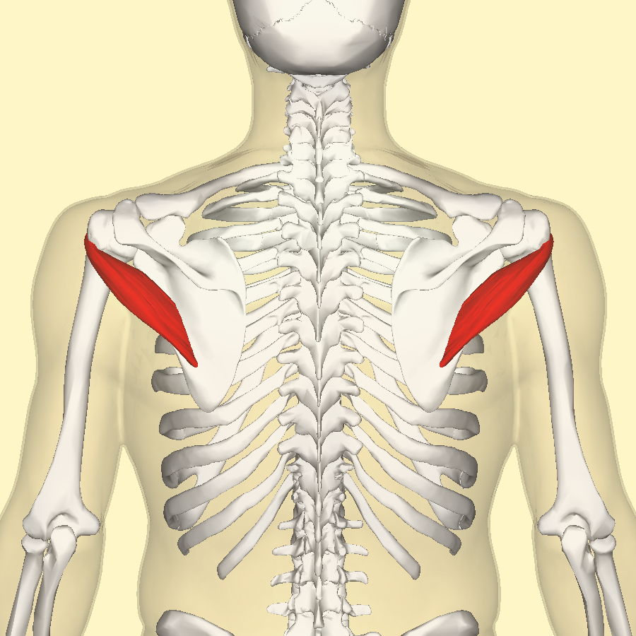
Teres Minor Muscle Wikipedia from upload.wikimedia.org The rotator cuff is a made up of four muscles in the shoulder, connecting the humerus to the scapula. The shoulder has about eight muscles that attach to the scapula, humerus, and clavicle. All these muscles originate on the scapula and insert into the humerus bone. For baseball pitchers, the teres minor demonstrates the highest level of emg activity of all the shoulder muscles during the. Start studying posterior shoulder muscles. You will hinder your progress if you overwork your shoulder muscles. Posterior and anterior muscles of the forearm forearm anatomy human anatomy picture upper limb anatomy. Each deltoid muscle has three heads, or distinct parts:
The clavicle (collarbone), the scapula (shoulder blade), and the humerus (upper arm bone) as well as associated muscles, ligaments and tendons.
Summary of the structure of the posterior shoulder muscles. Extends and laterally rotates the arm. Diagram shoulder muscles anatomy 101 shoulder muscles the handcare blog. The anterior (front) head, the lateral (middle/medial) head, and the posterior (rear) head. Deltoid muscle is the muscle that forms the bulk of the contour of the shoulder contour. • coracobrachialis • pectoralis major • subscapularis. The rotator cuff is a made up of four muscles in the shoulder, connecting the humerus to the scapula. The trapezius and underlying levator scapulae, rhomboideus. Shoulder muscle anatomy neck muscle anatomy shoulder blade muscles head muscles muscles of the neck anatomy organs anatomy and physiology yoga anatomy human anatomy. Muscles of the shoulder can be divided into two strata: Related online courses on physioplus. When a bilateral posterior dislocation is present, it is almost always secondary to seizure activity. You will hinder your progress if you overwork your shoulder muscles.
Nine muscles cross the shoulder joint. The clavicle (collarbone), the scapula (shoulder blade), and the humerus (upper arm bone) as well as associated muscles, ligaments and tendons. With seizure activity, the internal rotator muscles (teres major. Anterior graphic of the shoulder. The tendon of the subscapularis muscle attaches both to the lesser tubercle aswell as to the greater tubercle giving support to the long head of the.
Posterior Thorax And Shoulder Muscles Page 1 Line 17qq Com from img.17qq.com As a group, they are responsible for stabilizing the shoulder joint. Shoulder anatomy deltoid arm joint anterior brachialis coracobrachialis humerus medical. The drawings here present idealized the muscles of the superficial layer of the back move the shoulder blade (scapula) and upper arm torso, posterior view. The muscle of the anterior compartment (arm in anatomical position) function as flexors while the muscles of the posterior compartment function as extensors. The shoulder has about eight muscles that attach to the scapula, humerus, and clavicle. Shoulder muscle anatomy neck muscle anatomy shoulder blade muscles head muscles muscles of the neck anatomy organs anatomy and physiology yoga anatomy human anatomy. Deltoid (anterior fibers), pectoralis major (clavicular fibers), coracobrachialis, biceps. Posterior shoulder mobility deficits often lead to limitations in shoulder internal rotation and horizontal adduction.
Extends and laterally rotates the arm.
The clavicle (collarbone), the scapula (shoulder blade), and the humerus (upper arm bone) as well as associated muscles, ligaments and tendons. Anterior part of the deltoid: Posterior shoulder mobility deficits often lead to limitations in shoulder internal rotation and horizontal adduction. Summary of the structure of the posterior shoulder muscles. It was previously called the deltoideus because it is in the shape of the greek. The shoulder muscles are associated with movements of the upper limb. Right fibrous loop for intermediate digastric tendon. Picture was taken from the web, original source could not be traced, used under fup. Muscles of the shoulder can be divided into two strata: • coracobrachialis • pectoralis major • subscapularis. Want to learn more about it? Diagram shoulder muscles anatomy 101 shoulder muscles the handcare blog. The human shoulder is made up of three bones:
The shoulder anatomy includes the anterior, lateral & posterior deltoids, plus the rotator cuff. Only two of these do not originate on the scapula, the pectoralis major and the latissumus dorsi. The posterior muscles of the shoulder: Want to learn more about it? Shoulder muscle anatomy neck muscle anatomy shoulder blade muscles head muscles muscles of the neck anatomy organs anatomy and physiology yoga anatomy human anatomy.
Posterior Shoulder Muscles Diagram Quizlet from o.quizlet.com The rotator cuff is a made up of four muscles in the shoulder, connecting the humerus to the scapula. • coracobrachialis • pectoralis major • subscapularis. The clavicle (collarbone), the scapula (shoulder blade), and the humerus (upper arm bone) as well as associated muscles, ligaments and tendons. Related online courses on physioplus. As a group, they are responsible for stabilizing the shoulder joint. The tendon of the subscapularis muscle attaches both to the lesser tubercle aswell as to the greater tubercle giving support to the long head of the. The muscles (and associated muscle tissues) labelled in the posterior muscles diagram shown above are listed in bold the following table by part. With cross body stretching, people will often why is this relevant?
Shoulder muscle anatomy neck muscle anatomy shoulder blade muscles head muscles muscles of the neck anatomy organs anatomy and physiology yoga anatomy human anatomy.
Posterior and anterior muscles of the forearm forearm anatomy human anatomy picture upper limb anatomy. The shoulder muscles include skeletal muscles that are attached to the head of the humerus which performs various direct and indirect functions of the shoulder joints. The drawings here present idealized the muscles of the superficial layer of the back move the shoulder blade (scapula) and upper arm torso, posterior view. Flexes and medially rotates arm; As a group, they are responsible for stabilizing the shoulder joint. Posterior shoulder mobility deficits often lead to limitations in shoulder internal rotation and horizontal adduction. Posterior part of the deltoid: The clavicle (collarbone), the scapula (shoulder blade), and the humerus (upper arm bone) as well as associated muscles, ligaments and tendons. Learn their origins/insertions, functions & exercises. Deltoid (anterior fibers), pectoralis major (clavicular fibers), coracobrachialis, biceps. Published march 30, 2018 at 1300 × 910 in shoulder muscles diagrams. Extends and laterally rotates the arm. The deltoid (shoulder) muscle comprises three sections:
This muscle diagram is interactive: shoulder muscles diagram. As a group, they are responsible for stabilizing the shoulder joint.


0 Comments:
Posting Komentar